1: JAMA 1999 Nov 24;282(20):1942-6
CONTEXT: Diagnosis of infection with Borrelia burgdorferi, the cause of Lyme disease (LD), has been impeded by the lack of effective assays to detect active infection.
OBJECTIVE: To determine whether B. burgdorferi-specific immune complexes are detectable during active infection in LD.
DESIGN, SETTING, AND PATIENTS: Cross-sectional analysis of serum samples
from 168 patients fulfilling
Centers for Disease Control and Prevention surveillance criteria for
LD and 145 healthy and other disease controls conducted over 8 years. Tests
were performed blinded.
MAIN OUTCOME MEASURE: Detection of B. burgdorferi immune complexes by enzyme-linked immunosorbent assay and Western blot.
RESULTS: The B. burgdorferi immune complexes were found in
PMID: 10580460, UI: 20046405
Chronic fatigue syndrome (CFS) and Lyme disease often share clinical features, especially fatigue, contributing to concern that Borrelia burgdorferi (Bb), the cause of Lyme disease, may underlie CFS symptoms. We examined
PMID: 10522896, UI: 99450731
3: Wien Klin Wochenschr 1999 May 7;111(9):368-70
Ceftriaxone associated hemolysis.
Maraspin V, Lotric-Furlan S, Strle F
Department of Infectious Diseases, University Medical
Centre Ljubljana,
Slovenia.
A 48-year-old immunocompetent women treated with ceftriaxone
2 g daily i.v. for
late Lyme borreliosis developed severe haemolytic anaemia.
The patient had
previously received the same antibiotic two times without
any side effects. The
first clinical signs began to appear on the 7th day of
treatment. The
patient developed severe anaemia with a haemoglobin level
of 45 mg/l on day 10;
thereafter she ceased to receive the antibiotic. The
outcome was favourable. The
clinical course and serologic results suggest that severe
anaemia was induced by
ceftriaxone and that drug adsorption as well as immune
complex mechanisms were
involved in the pathogenesis.
PMID: 10407998, UI: 99336336
4: Med Dosw Mikrobiol 1998;50(1-2):97-103
Binding of antibodies specific to Borrelia burgdorferi in circulating immune complexes can lead to false negative results in serological tests. The aim of our study was to determine the presence of IgM antibodies to Borrelia burgdorferi bound in immune complexes in 52 sera of foresters the National Park in Karkonosze.
Publication Types:
Clinical trial
PMID: 9857619, UI: 99074946
5: J Immunol Methods 1998 Sep 1;218(1-2):9-17

A one-step immunoassay for simultaneous detection of serum IgG and IgM antibodies to Borrelia burgdorferi has been developed.
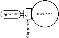
The assay is based on C1q, which binds to immune complexes containing IgG and/or IgM antibodies (1, 2).
Micro-beads pre-coated with antibodies to human C1q are mixed with human serum samples and fluorochrome-labelled B. burgdorferi flagellum antigen. In the presence of serum IgG and/or IgM antibodies to B. burgdorferi, fluorochrome-labelled antigen/antibody complexes are formed. These are then bound by serum C1q and are subsequently captured on the anti-C1q-coated beads.
The sample is analysed on a flow cytometer and the presence of fluorescent beads is, thus, indicative of a positive test result. In the present study the sensitivity and specificity of the assay are compared to those of the indirect IDEIA B. burgdorferi IgG and the mu-chain capture IDEIA B. burgdorferi IgM ELISAs for separate determination of IgG and IgM.
Detection using a flow cytometer can be performed without separation of the beads from the reaction mixture, which means that, in practice, the method is carried out as a one-step assay and it is, thus, very suitable for automation. Other advantages of this kind of assay includes an antibody/antigen reaction which occurs in solution and the potential of using the method for the detection of antibodies against several antigens from the same or different infectious agents (multi-parameter screening).
PMID: 9819119, UI: 99034428
Abstract edited for enhanced legibility by J. Gruber

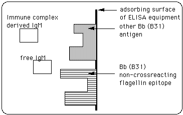
We previously reported on the efficacy of the enzyme-linked IgM capture immune complex (IC) biotinylated antigen assay (EMIBA) for the seroconfirmation of early Lyme disease and active infection with Borrelia burgdorferi (enhanced efficacy due to elimination of competition between immunglobulins and other components for binding sites on antigen, J. Gruber). In earlier work we identified non-cross-reacting epitopes of a number of B. burgdorferi proteins, including flagellin.
We now report on an improvement in the performance of EMIBA with the addition of a biotinylated form of a synthetic non-cross-reacting immunodominant flagellin peptide to the biotinylated B. burgdorferi B31 sonicate antigen source with the avidin-biotinylated peroxidase complex detection system used in our recently developed indirect IgM-capture immune complex-based assay (EMIBA).
As in our previous studies, we compared
PMID: 9542940, UI: 98201939
The present recommendation for serologic confirmation of Lyme disease (LD) calls for immunoblotting in support of positive or equivocal ELISA. Borrelia burgdorferi releases large quantities of proteins, suggesting that specific antibodies in serum might be trapped in immune complexes (ICs), rendering the antibodies undetectable by standard assays using unmodified serum. Production of ICs requires ongoing antigen production, so persistence of IC might be a marker of ongoing or persisting infection. We developed an immunoglobulin M (IgM) capture assay (EMIBA) measuring IC-derived IgM antibodies and tested it using three well-defined LD populations from
PMID: 11526153 [PubMed - indexed for MEDLINE]
7: Przegl Epidemiol 1997;51(4):451-5
[Changes in granulocytic receptors for FcR IgG and CR with circulating immune complexes in patients with lyme borreliosis].
[Article in Polish]
Izycka A, Jablonska E, Pancewicz S, Zajkowska J, Swierzbinska R, Kondrusik M, Izycki T
Zaklad Immunopatologii Akademii Medycznej w Bialymstoku.
This study aimed to estimate some PMN functions, involving phagocytic
activity
in patients with Lyme borreliosis. Decreased percentage PMN with FcR
and CR
receptors was observed. Increased immune complexes levels in the serum
of
patients before, and their normalization after treatment were found.
These
results indicate a depression of non-specific cellular response, which
can
influence the general immune system in patients with Lyme borreliosis.
PMID: 9562795, UI: 98223888
To investigate whether circulating immune complexes can be used as a
disease marker for assessment of the activity of Lyme disease and for monitoring
patients response to treatment, we tested 104 sera from patients with different
stages of Lyme disease using the C1q
enzyme-linked immunosorbent assay (ELISA) and a modified Raji cell test.
PMID: 9403844, UI: 98067621
Serological assays for detection of canine antibodies to the Lyme agent generally have been difficult to validate because an acceptable standard of comparison such as unequivocal proof of infection status has not been available. For practical and logistical reasons, it has not been possible to use culture of organism from infected animals, seroconversion in a large number of field dogs, or clinical criteria as the standard of comparison for validation of assays. Therefore, estimates of diagnostic sensitivity and specificity based on an appropriate gold standard have not been available. When it was discovered how to infect laboratory dogs via ticks infected with Borrelia burgdorferi, it was possible to define the kinetics and magnitude of the antibody response that might be expected in nature. ELISA and Western immunoblot data from experimental dogs were then compared and correlated with results of the same tests on dogs from endemic and nonendomic areas. Coupled with studies on cross-reactive antibodies elicited from other infectious agents or autoimmune phenomena, it was possible to account for interfering antibodies and to establish estimates of diagnostic sensitivity and specificity for the ELISA based on objective criteria. Such validated assays can predict, with a relatively high degree of proficiency, the infection and/or vaccinal status of animals. These assays have shown that some dogs, vaccinated with the commercially available whole-cell Lyme bacterins develop typical signs of Lyme disease but have no evidence of an underlying infection; antibody elicited only by the vaccine and not by infection is detectable in these animals. Western immunoblot can also confirm infection in animals of equivocal ELISA status if their bands have been evaluated for specificity of antibodies to B burgdorferi. Serology can be a very useful aid in the diagnosis of Lyme disease, but it requires that the assays used have been subjected to rigorous validation criteria. When that is not performed, an unacceptable level of false-positive and false-negative test results is virtually assured.
NLM PUBMED CIT. ID: 8942214 NLM CIT. ID: 97097649
9: Przegl Epidemiol 1996;50(3):253-7
[Immunologic reaction in patients with arthritis in the course of borreliosis
with Lyme disease].
[Article in Polish]
Flisiak R, Wiercinska-Drapalo A, Prokopowicz D
Klinika Obserwacyjno-Zakazna Akademii Medycznej w Bialymstoku.
Cell mediated as well as humoral immune response was evaluated in 14
lyme
arthritis patients. Significant decrease of T cells percentage was
observed in
comparison to normal values. It was related particularly to CD4+ subset
and
resulted in significant decrease of CD4+/CD8+ ratio. Statistically
significant
increase of B cells percentage was accompanied by elevated concentrations
of
immunoglobulin M, complement components C3 and C4. Immunoglobulins
A and G, as
well as circulating immune complexes remained on the normal level.
These results
indicate suppression of cellular and activation of humoral immune response
in
patients with Lyme arthritis.
PMID: 8927735, UI: 97027669
OBJECTIVE:
To determine the potential of detection in CSF of specific Borrelia
burgdorferi antigen, OspA, as a marker of infection in neurologic Lyme
disease and compare this with the detection of antibody.
DESIGN:
CSF from 83 neurologic patients in an area highly endemic for Lyme
disease was examined prospectively for
NLM PUBMED CIT. ID: 7501150 NLM CIT. ID: 96063525
The sensitivities and specificities of three enzyme-linked immunosorbent assays (ELISAs) for Borrelia burgdorferi antibodies were compared for 41 patients presenting with symptoms compatible with late Lyme borreliosis (LB) and 37 healthy controls. All subjects were living in southwestern Finland, where LB is endemic. Only patients with culture- or PCR-proven disease were enrolled in the study.
The antigens of the ELISAs consisted of
CONCLUSIONS:
These results show that antibodies to B. burgdorferi may be present
in low levels or even absent in patients with culture- or PCR-proven late
LB. Therefore, in addition to serological testing, the use of PCR and cultivation
is recommended in the diagnosis of LB.
NLM PUBMED CIT. ID: 7494012 NLM CIT. ID: 96025135
COMMENTS:
Comment in: Am J Ophthalmol 1995 Aug;120(2):263-4
PURPOSE:
To establish a diagnosis, in a group of patients we studied the characteristics
of ocular Lyme borreliosis.
METHODS:
During a two-year period, 236 patients with prolonged external ocular
inflammation, uveitis, retinitis, optic neuritis, or unexplained neuro-ophthalmic
symptoms were examined for Lyme borreliosis. Antibodies to Borrelia burgdorferi
were measured by indirect ELISA and western blot. Cerebrospinal fluid was
also analyzed by polymerase chain reaction.
RESULTS:
CONCLUSIONS:
Late-phase ocular Lyme borreliosis is probably underdiagnosed because
of weak seropositivity or seronegativity in ELISA assays. Ocular borrelial
manifestations show characteristics resembling those seen in syphilis.
NLM PUBMED CIT. ID: 7832219 NLM CIT. ID: 95133613
10: Lab Invest 1995 Feb;72(2):146-60
Chronic lyme disease in the rhesus monkey.
Roberts ED, Bohm RP Jr, Cogswell FB, Lanners HN, Lowrie RC Jr, Povinelli
L,
Piesman J, Philipp MT
Department of Pathology, Tulane Regional Primate Research Center, Tulane
University Medical Center, Covington, Louisiana.
BACKGROUND: We have previously reported the clinical, pathologic, and
immunologic features of "early" Borrelia burgdorferi infection in rhesus
monkeys
(3). We have now evaluated these features during the chronic phase
of Lyme
disease in this animal model.
EXPERIMENTAL DESIGN: Clinical signs, and pathologic changes at the gross
and microscopic levels, were investigated 6 months post-infection in several
organ systems of five rhesus macaques (Macaca mulatta), which were infected
with Borrelia burgdorferi by allowing infected Ixodes scapularis nymphal
ticks to feed on them. A sixth animal was used as an
uninfected control. Borrelia antigens recognized by serum antibody
were identified longitudinally by Western blot analysis, and C1q-binding
immune complexes were quantified. Localization of the spirochete in the
tissues was achieved by immunohistochemistry and in vitro culture. The
species of spirocheta cultured was confirmed by the polymerase chain reaction.
RESULTS: Chronic arthritis was observed in five out of five animals. The knee and elbow joints were the most consistently affected. Articular cartilage necrosis and/or degenerative arthropathy were the most severe joint structural changes. Synovial cell hyperplasia and a mononuclear/lymphocyte infiltrate were commonly seen. Nerve lesions were also observed, including nerve sheath fibrosis and focal demyelinization of the spinal cord. Peripheral neuropathy was observed in five out of five animals and could be correlated in the most severely affected monkey with the presence of higher levels of circulating immune complexes. Differences in disease severity did not correlate with differences in the antigens recognized on Western blot analysis.
CONCLUSIONS: B. burgdorferi infection in rhesus macaques mirrors several aspects of both the early and chronic phases of the disease in humans. This animal model will facilitate the study of the pathogenesis of Lyme arthritis and neuroborreliosis.
Comments:
Comment in: Lab Invest 1995 Feb;72(2):127-30
PMID: 7853849, UI: 95156954
11: J Infect Dis 1994 Oct;170(4):890-3
Fc- and non-Fc-mediated phagocytosis of Borrelia burgdorferi by macrophages.
Montgomery RR, Nathanson MH, Malawista SE
Department of Internal Medicine, Yale University School of Medicine,
New Haven,
Connecticut 06520.
The Fc receptor (FcR) for immunoglobulin has been assigned a major role
in the
ingestion of Borrelia burgdorferi, the Lyme disease spirochete, by
macrophages.
Yet macrophages readily take up and kill B. burgdorferi that have not
been
opsonized. By use of doubly-labeled macrophages infected with spirochetes
and
analyzed by confocal fluorescence microscopy, simultaneous localization
of both
FcR and spirochetes (opsonized and unopsonized) was quantified. After
infection
with unopsonized spirochetes, bacterial surface antigen and the FcR
remained
distinct, confirming the expectation that unopsonized uptake of B.
burgdorferi
is largely independent of the FcR. A similar lack of colocalization
was seen
when opsonized spirochetes were ingested by macrophages whose FcRs
were
sequestered by an immune complex-coated substrate. Furthermore, comparable
efficiency of uptake was observed whether or not the FcR was engaged.
PMID: 7930732, UI: 95016039
Borrelia burgdorferi (Bb), the cause of Lyme disease, has appeared not to evoke a detectable specific antibody response in humans until long after infection.
This delayed response has been a biologic puzzle and has hampered early diagnosis.
PMID: 8040289, UI: 94314934
13: Pediatr Res 1993 Jan;33(1 Suppl):S90-4
Pathogenesis of immune-mediated neuropathies.
Rostami AM
Department of Neurology, University of Pennsylvania School of Medicine,
Philadelphia 19104.
A variety of peripheral neuropathies are believed to be immune-mediated.
Acute
inflammatory demyelinating polyneuropathy or Guillain-Barre syndrome
(GBS) is
the prototype of these neuropathies. GBS is characterized by acute
progressive
motor weakness of the extremities and of bulbar and facial musculature.
Deep
tendon reflexes are reduced or absent, and sensory symptoms are mild.
Respiratory failure and autonomic dysfunction may be seen. The cerebrospinal
fluid shows increased protein and no or very few cells. The nerve conduction
velocity is slowed, and the pathology shows segmental demyelination
with
mononuclear cell infiltration. Studies from man and experimental animals
suggest
an immunologic basis for demyelination of the peripheral nerves in
GBS, but the
mechanism is not well understood.
Experimental allergic neuritis, an animal
model of GBS, is induced in laboratory animals by immunization with
myelin P2
protein, some peptides of P2 protein, and galactocerebroside. The animals
develop weakness and show electrophysiologic and pathologic features
similar to
GBS. P2-reactive T cells and antigalactocerebroside antisera can adoptively
transfer experimental allergic neuritis. Various antibodies to peripheral
nerve
myelin and circulating immune complexes have been found in patients
with GBS.
The target antigen(s) for these antibodies are not well understood,
but neutral
glycolipids cross-reactive with Forssman antigen and gangliosides are
possible
candidates.
The mainstay of therapy is the management of the paralyzed patient.
Steroids are ineffective. Plasmapheresis, especially early in the course
of the
disease, can shorten the duration of paralysis and intubation. Results
from a
multicenter study in the Netherlands demonstrate the efficacy of high-dose
immune globulin therapy in GBS.
Publication Types:
Review
Review, tutorial
PMID: 8381954, UI: 93165407
NLM PUBMED CIT. ID: 1583267 NLM CIT. ID: 92259900
J Infect Dis 1991 Feb;163(2):305-10
A past history of clinical Lyme borreliosis and the 6-month incidence of clinical and asymptomatic Lyme borreliosis was studied prospectively in a high-risk population. In the spring, blood samples were drawn from 950 Swiss orienteers, who also answered a questionnaire. IgG anti-Borrelia burgdorferi antibodies were detected by ELISA. Positive IgG antibodies were seen in 248 (26.1%), in contrast to 3.9%-6.0% in two groups of controls (n = 101). Of the orienteers, 1.9%-3.1% had a past history of definite or probable clinical Lyme borreliosis. Six months later a second blood sample was obtained from 755 participants, 558 (73.9%) of whom were seronegative initially; 45 (8.1%) had seroconverted from negative to positive. Only 1 (2.2%) developed clinical Lyme borreliosis. Among all participants, the 6-month incidence of clinical Lyme borreliosis was 0.8% (6/755) but was much higher (8.1%) for asymptomatic seroconversion (45/558). In conclusion, positive Lyme serology was common in Swiss orienteers, but clinical disease occurred infrequently.
NLM PUBMED CIT. ID: 1988513 NLM CIT. ID: 91108157
14: Ann Ital Med Int 1991 Oct-Dec;6(4 Pt 2):483-90
Collagenopathic cardiopathies.
Carcassi U, Passiu G
II Cattedra di Reumatologia, Universita degli Studi di Roma La Sapienza, Italy.
Collagenopathic cardiopathies are a subject of extreme etiologic, pathogenetic
and clinical interest. These disorders are associated with congenital
or
acquired anomalies of the connective tissue and because of the diffusion
and
nearly total distribution of this tissue, have a higher frequency than
what has
been previously estimated. The collagenopathic cardiopathies, can be
divided
into two main groups: one deriving from hereditary connective tissue
diseases,
and the other from acquired connective tissue diseases. The first group
has a
Mendelian type of transmission whereas the other appears to be secondary
to
various kinds of stimuli (viral, immunologic etc.) although polygenic
factors
are present. Of the first group we considered Marfan's syndrome, the
Ehlers-Danlos syndrome, osteogenesis imperfecta, pseudoxanthoma elasticum,
cutis
laxa and the diseases of the fundamental substance with particular
reference to
mucopolysaccharidosis type 1H (Hurler's syndrome). In all of these
disorders a
specific metabolic disturbance is responsible for the cardiovascular
damage
which is expressed, depending on the specific genetic component in
a more or
less serious form. Among the acquired diseases of the connective tissue,
we
examined rheumatoid arthritis, systemic lupus erythematosus,
polydermatomyositis, scleroderma; of the reactive arthritis, rheumatic
fever; of
the seronegative forms, spondyloarthritis, ankylosing spondylitis and
Reiter's
syndrome, mixed connective tissue disease and Lyme's disease. It must
be
emphasized that all of these disorders share relatively common pathogenetic
characteristics which point to the importance of the presence of various
types
of antigens, immune complexes and the significant role of some of the
histocompatibility antigens, as well as possible disturbances of cell-mediated
immunity.
PMID: 1840815, UI: 93002044
15: Cutis 1991 Apr;47(4):229-30, 232
Diagnosing Lyme disease: often simple, often difficult.
Schutzer SE, Schwartz RA
Department of Medicine, UMDNJ-New Jersey Medical School, Newark 07103.
Lyme disease has as its hallmark erythema migrans. However, it is only
present
in about one half of the patients who contract this disease. In its
absence, the
diagnosis of Lyme disease may be difficult. It depends upon a compatible
history
of exposure and clinical signs and symptoms together with positive
results of
serologic testing. Unfortunately, seronegativity for antibody to the
pathogen
may occur both during the first six weeks of infection and be chronic
due to the
reactive antibody being bound in immune complexes. The selective use
of new
diagnostic tests may be required to confirm the diagnosis. These tests
include
assays for antibody or antigen analysis of immune complex components,
as well as
polymerase chain reactions.
Publication Types:
Review
Review, tutorial
PMID: 2070642, UI: 91300888
We analyzed cerebrospinal fluid (CSF) from 32 patients with neurological symptoms and evidence of Borrelia burgdorferi infection
PMID: 2285261, UI: 91136153
To find out whether apparent seronegativity in patients strongly suspected
of having Lyme disease can be due to sequestration of antibodies in immune
complexes, such complexes were isolated and tested for antibody to Borrelia
burgdorferi.
PMID: 1967770, UI: 90135740
Acquired transient autoimmune reactions in Lyme arthritis: correlation between rheumatoid factor and disease activity.
Goebel KM, Krause A, Neurath F
Department of Medicine, University Hospital, Marburg, West Germany.
Lyme spirochaetal disease (LSD) is a complex multisystem disorder which has been recognized as a separate entity due to its close geographic clustering of affected patients. The study aimed at evaluating the clinical and immunological features of LSD with chronic symptoms of meningoradiculitis, carditis and pauciarticular arthritis. Six patients with LSD and erosive arthritis who developed an increase of serum IgM rheumatoid factor (RF) which correlated with the inflammatory activity of the disease are described in detail. Besides raised IgG antibody titers to Borrelia burgdorferi (B. burgd.) antigen measured by ELISA technique, circulating immune complexes, antinuclear antibodies (ANA) and RF measured by laser nephelometric immunoassay were detected. Increased ANA and RF antibody rates suggest that LSD may closely be linked with transient autoimmune phenomena. Thus, in some cases, B. burgd. antigens might be able to produce a strong polyclonal B-cell stimulation, hence leading to an unspecific autoimmune reaction. But the question remains if transient unspecific autoimmune reactions actually take part in the pathogenesis of LSD.
PMID: 3238365, UI: 89186685
19: Med Clin North Am 1985 Jul;69(4):623-36
Publication Types:
Review
PMID: 3903373, UI: 86039054
20: Infection 1985 May-Jun;13(3):156
Immune complexes in leptospirosis.
Galli M, Esposito R, Crocchiolo P, Chemotti M, Gasparro M, Dall'Aglio PP
Publication Types:
Letter
PMID: 4030109, UI: 85287599
21: Acta Paediatr Scand 1985 Jan;74(1):133-6
Lyme disease in a 12-year-old girl.
Sturfelt G, Cavell B
We report the case of a 12-year-old girl with erythema chronicum migrans,
aseptic meningitis and knee arthralgia. Rise of specific antibody titre
against
an Ixodes ricinus spirochaete was demonstrated. Circulating immune
complexes and
high levels of C1r-C1s-C1IA complexes indicating activation of the
complement
system via the classical pathway were found. The clinical features
and the
laboratory findings warranted a diagnosis of Lyme disease.
PMID: 3984718, UI: 85171163
22: Adv Clin Chem 1985;24:1-60
Immune complexes in man: detection and clinical significance.
McDougal JS, McDuffie FC
Publication Types:
Review
PMID: 2936066, UI: 86126364
23: J Infect Dis 1984 Oct;150(4):497-507
Interactions of phagocytes with the Lyme disease spirochete: role of
the Fc
receptor.
Benach JL, Fleit HB, Habicht GS, Coleman JL, Bosler EM, Lane BP
The phagocytic capacity of murine and human mononuclear and polymorphonuclear
phagocytes (including peripheral blood monocytes and neutrophils),
rabbit and
murine peritoneal exudate cells, and the murine macrophage cell line
P388D1
against the Lyme disease spirochete was studied. All of these cells
were capable
of phagocytosing the spirochete; phagocytosis was measured by the uptake
of
radiolabeled spirochetes, the appearance of immunofluorescent bodies
in
phagocytic cells, and electron microscopy. Both opsonized and nonopsonized
organisms were phagocytosed. The uptake of opsonized organisms by neutrophils
was blocked by a monoclonal antibody specific for the Fc receptor and
by immune
complexes; these findings suggested that most phagocytosis is mediated
by the Fc
receptor. Similarly, the uptake of opsonized organisms by human monocytes
was
inhibited by human monomeric IgG1 and by immune complexes. These results
illustrate the role of immune phagocytosis of spirochetes in host defense
against Lyme disease.
PMID: 6386995, UI: 85031981
24: Yale J Biol Med 1984 Jul-Aug;57(4):589-93
Circulating C1q binding material was found in nearly all patients at onset of erythema chronicum migrans, the skin lesion that marks the onset of infection with the causative spirochete. In patients with only subsequent arthritis this material tended to localize to joints where it gradually increased in concentrations with greater duration of joint inflammation. In joints, its concentration correlated positively with the number of synovial fluid polymorphonuclear leukocytes. Despite the prolonged presence of putative immune complexes, rheumatoid factors could not be demonstrated.
These observations suggest that phlogistic immune complexes based on spirochete antigens form locally within joints during chronic Lyme arthritis.
PMID: 6334939, UI: 85092791
25: Semin Arthritis Rheum 1984 Feb;13(3):229-34
Despite systemic clearing in some patients, the immune complexes localize to the joints where a chronic synovitis develops, similar to rheumatoid arthritis. Why the immune complexes localize to the joints is an enigma. It is tempting to postulate that this localization occurs because of an altered immune response in a genetically predisposed group. However, three of 10 patients with chronic arthritis did not have the B-cell alloantigen DRw2.
PMID: 6233699, UI: 84223994
26: Clin Exp Rheumatol 1983 Oct-Dec;1(4):327-32
Saturable, high-avidity monocyte receptors for monomeric IgG and Fc
fragments
increase in SLE and lyme disease.
Hardin JA, Downs JT
We have devised an assay for quantifying high-avidity Fc receptors for
monomeric
IgG on peripheral blood monocytes. In the development of a radiolabelled
ligand
for the assay, we found that Fc fragments offer several advantages
over 7S-IgG.
Compared to the latter ligand, the fragments interacted more cleanly
with a
single high-avidity binding site, appeared to have easier access to
this site,
and, since they showed no binding to Millipore filters, their use made
possible
a wash procedure that was convenient and rapid, thus minimizing loss
of
specifically bound ligand. Application of the assay to a study of ten
normal
controls, five patients with SLE, and three patients with Lyme disease
demonstrated that normal monocytes bear approximately 10,000 high-avidity
binding sites per cell. In contrast, patient monocytes bore significantly
more
Fc receptors; on average their cells had about 40,000 such sites per
cell (P =
0.01) and sometimes as many as 100,000 sites per cell. Both normal
and patient
monocytes bound IgG or Fc fragments with an apparent association constant
(KA)
of approximately 10(8) M-1. The majority of patients with active SLE
and Lyme
disease had serum C1q-binding material compatible with the presence
of
circulating immune complexes. This study shows that these putative
circulating
immune complexes do not necessarily lead to a reduction in the number
of Fc
receptors on peripheral blood monocytes. Rather, the data suggest that
in the
course of immune mediated diseases, either monocytes are activated
in vivo to
express greater numbers of Fc receptors, or a subset of monocytes bearing
more
Fc receptors is expanded.
PMID: 6241857, UI: 85177804
27: Hum Pathol 1983 Apr;14(4):343-9
Immune complex assays in rheumatic diseases.
Agnello V
A wide variety of antigen-nonspecific immune complex assays have been
developed
in recent years for the detection and quantitation of immune complexes
in
pathologic fluids. These assays detect complexed antibody regardless
of the
antigen involved. Almost all of these assays use biologic reagents
that may
react with substances other than complexed antibody. In addition, the
assays do
not differentiate nonspecifically aggregated antibody from antigen-complexed
antibody. Hence, these assays are not absolute tests for immune complexes.
On
the basis of studies using these assays, "immune complexes" have been
detected
in a large number of rheumatic diseases. While these findings have
been of
considerable investigative interest, thus far they have been of little
practical
clinical utility. The detection of immune complexes has not been shown
to be
essential in any clinical conditions but may be helpful in monitoring
disease
activity in systemic lupus erythematosus (SLE) and may provide useful
diagnostic
information in two rare syndromes, Lyme arthritis and SLE-related syndrome.
Publication Types:
Review
PMID: 6187658, UI: 83159380
28: Pediatr Infect Dis 1983 Jan-Feb;2(1):47-9
Lyme disease with neurologic abnormalities.
Darras BT, Annunziato D, Leggiadro RJ
PMID: 6220264, UI: 83169227
29: Nouv Presse Med 1982 Jan 9;11(1):39-41
[Lyme's disease. A clinical case observed in Western France].
[Article in French]
Mallecourt J, Landureau M, Wirth AM
The clinical story of a young woman with chronic erythema migrans followed
by
polyradiculoneuritis and recurrent oligoarthritis is reported. The
story
corresponds to the disease described by Steere et al. [7, 8, 9, 10]
in 1976 and
known in the U.S.A. as "Lyme's disease". The condition is epidemic
and occurs
during the summer. It begins with skin lesions characteristic of chronic
erythema migrans, which are consecutive to tick bite. This is followed,
a few
days or weeks later, by neurological disorders (aseptic meningitis,
encephalitis, cranial nerve paralysis and/or polyradiculoneuritis),
transient
and recurrent attacks or arthritis mostly in the larger joints and,
occasionally, conduction disorders in the heart. The course of the
disease is
that of an inflammatory condition. The presence of immune complexes
in the serum
and synovial fluid is suggestive of local and systemic immune reaction
to a
hypothetically viral agent introduced by the tick bite. The fact that
the
incidence of DR W2 antigen is greater in patients with severe lesions
suggests
individual predisposition.
PMID: 7058122, UI: 82126736
30: Dtsch Med Wochenschr 1980 Dec 19;105(51):1779-81
[Erythema chronicum migrans with arthritis].
[Article in German]
Ackermann R, Runne U, Klenk W, Dienst C
In a 46-year-old woman arthritis developed in several large joints eight
weeks
after the onset of erythema chronicum migrans. The joints of the leg
were
swollen and painful. In addition there was painful involvement bilaterally
of
knee, hip and elbow joints. Circulating immune complexes were demonstrated
in
serum and the erythrocyte sedimentation rate was moderately increased.
All other
laboratory and radiological tests were within normal limits. On symptomatic
treatment the arthritis regressed without sequelae within six weeks.
The disease
is nosologically related to Lyme disease recently described in the
North East of
the U.S.A. in which erythema chronicum migrans is followed by arthritis;
here,
too, circulating immune complexes have been demonstrated.
PMID: 7439072, UI: 81066455
31: N Engl J Med 1979 Dec 20;301(25):1358-63
Immune complexes and the evolution of Lyme arthritis. Dissemination
and
localization of abnormal C1q binding activity.
Hardin JA, Steere AC, Malawista SE
In a prospective study of 78 patients with Lyme arthritis, abnormal
serum C1q
binding activity was present at the initial onset of erythema chronicum
migrans
in nearly all cases. The abnormal binding persisted in patients with
subsequent
nerve or heart involvement. In contrast, among those with only subsequent
arthritis, it usually disappeared within three months (P = 0.018).
However, in
the synovial fluid of affected joints, abnormal binding was uniformly
present,
and always to a greater extent than in the circulation. The abnormally
reactive
material behaved like antigen-antibody complexes. It had a density
of 19S or
greater, dissociated below pH 4.2, and lacked antiglobulin activity.
Cryoprecipitates containing immunoglobulin were good but insensitive
predictors
of its presence, but immune complexes themselves did not seem primarily
responsible for cryoprecipitability. Thus, as judged by C1q binding,
immune
complexes remain disseminated in certain patients with Lyme arthritis
but
localize to joints in others.
PMID: 503166, UI: 80054658
32: J Clin Invest 1979 Mar;63(3):468-77
Circulating immune complexes in Lyme arthritis. Detection by the 125I-C1q
binding, C1q solid phase, and Raji cell assays.
Hardin JA, Walker LC, Steere AC, Trumble TC, Tung KS, Williams RC Jr,
Ruddy S,
Malawista SE
PMID: 429566, UI: 79151567
33: Am Fam Physician 1978 Jan;17(1):161-6
Arthralgias and arthritis in viral infections.
Franklin EC
Arthritis and arthralgias are common in many viral infections. They
are
particularly prominent with hepatitis B virus and rubella infection,
where they
may be the major presenting symptom. "Lyme arthritis" is also associated
with a
virus. Similar symptoms are occasionally seen with adenovirus, Coxsackie
and
echovirus infection. Joint lesions are due to the deposition of immune
complexes
and not direct viral infection. While the arthritis is usually transient
and
self-limited, the physician must consider viral infections as etiologic
agents
of arthralgias and arthritis.
PMID: 146421, UI: 78100292
34: Science 1977 Jun 3;196(4294):1121-2
Erythema chronicum migrans and Lyme arthritis: cryoimmunoglobulins and
clinical
activity of skin and joints.
Steere AC, Hardin JA, Malawista SE
We report the presence of serum cryoimmunoglobulins in patients with
attacks of
a newly described epidemic arthritis--Lyme arthritis--and in some patients
with
a characteristic skin lesion--erythema chronicum migrans--that sometimes
precedes the onset of the arthritis. Seven patients who had cryoimmunoglobulins
at the time of the skin lesion have developed arthritis; four patients
without
them have not. The cryoglobulins in patients with the skin lesion consisted
primarily of immunoglobulin M (IgM); those in patients with arthritis
often
included both IgM and IgG. These findings support the hypothesis that
a common
origin exists for the skin and joint lesions and suggest that circulating
immune
complexes may have a pathogenetic role in Lyme arthritis.
PMID: 870973, UI: 77174694